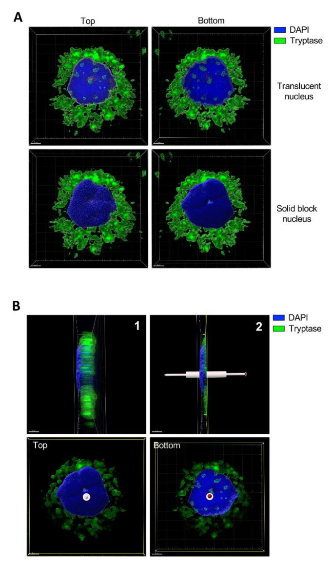
Figure 6.
Tryptase is found in the nuclei of primary human skin mast cells. Human skin mast cells were stained for tryptase and with a nuclear dye (Hoechst33342), followed by confocal microscopy analysis. (A) 3-D view generated from Z-stack sections of a representative mast cell. Upper panels are showing top and bottom views from the same cell with a translucent nuclear structure. Note the presence of tryptase staining within the nucleus and that the staining is concealed when switching to solid block (lower panels), revealing that the tryptase is present within the nuclear compartment. Abundant tryptase staining is seen in the cytoplasm, representing secretory granules. (B) 1: Right side view from A (lower right panel). 2: Cross-section cut dividing the nucleus in half. Lower panels are showing top and bottom views from panel B2. Note that tryptase is present in different layers of the nucleus. The staining was performed twice and panels are showing a representative cell from more than ten individual cells analyzed.

