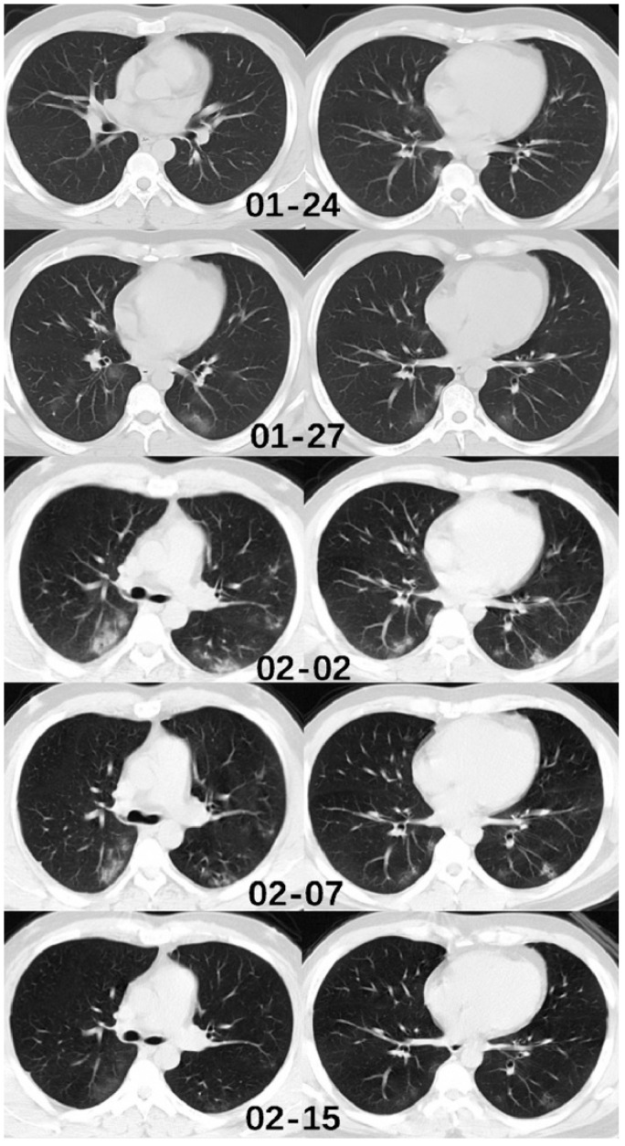
Figure 3.
Chest CT of a 37-year-old male patient. This individual returned from Wuhan to Wenzhou on January 19 and inflicted with cough and expectoration. The first chest CT was conducted on January 24, which showed subtle peripheral ground-glass opacity in the middle lobe, and right inferior lobe. The second CT images, of January 27, and the third examination, on February 2, showed a significant increase of lesion numbers and density, especially in both lower lobes. The fourth CT images, of February 7, showed a decrease in the density of the pulmonary lesions. On the fifth CT examination of February 15, the lesions were absorbed, and the patient was discharged [52]. Reprinted (adapted) with permission from Elsevier.

