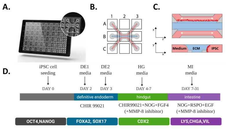
Figure 1.
Seeding and differentiation of hiPSC in the 3-lane OrganoPlate. (A) Artist’s impression of the bottom-side of the 3-lane OrganoPlate containing 40 individual microfluidic chips. The inlay shows an individual chip that is communicating with nine connecting wells. (B) Scheme of a single chip containing three microfluidic channels all having top, middle and bottom inlets (A1, B1 and C1), outlets (A3, B3 and C3) and an observation window (B2). (C) A schematic representation of hiPSC introduction to a microfluidic chip in a top (xz plane) and cross sectional view (yz plane); single iPSCs (in red) are seeded in the top channel and are allowed to adhere for up to 24 h after which media is added and perfusion initiated, cells start to form tubules by first growing against the ECM gel (blue) and then covering the whole channel. (D) Schematic of workflow for directed on plate differentiation of iPSC towards gut tubules.

