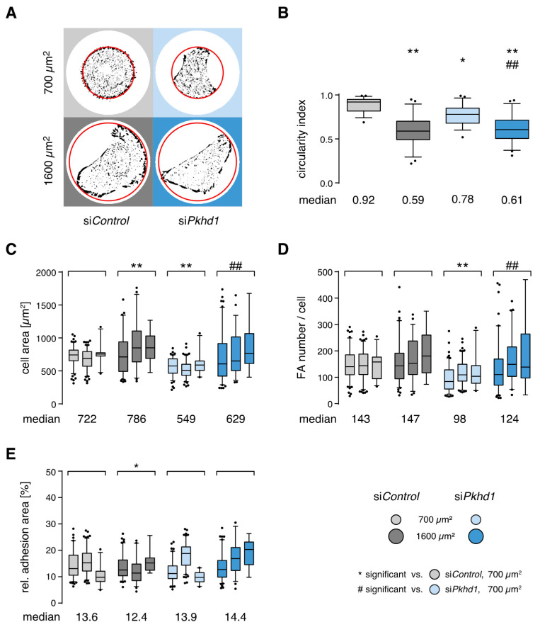
Figure 2.
Effects of FPC deficiency and confinement on cell shape and adhesion sites. (A) Representative cell shape and adhesion sites of siRNA-treated cells. Four hours after seeding onto collagen-coated micropatterns, cells were fixed and stained for vinculin to mark cell–ECM adhesions. Red line indicates the outline of disk-shaped adhesion area; diameter size: 30 and 45 µm for 700 and 1600 µm2 pattern, respectively. (B) Cell geometry determined by relation of short to long cell axis (circularity index). All box plots with whiskers 5/95% (n = 50 cells per condition, two-way ANOVA/Tukey’s). (C–E) Spreading area, number of adhesion sites per cell and relative adhesion area extracted from fluorescence images of steady-state adhesion for FPC-deficient cells and controls in spheroid culture conditions. (n = 3 independent experiments, >250/170 cells per condition for 700/1600 µm2; two-way ANOVA/Tukey’s, p < 0.05/0.01, */** or #/##.)

