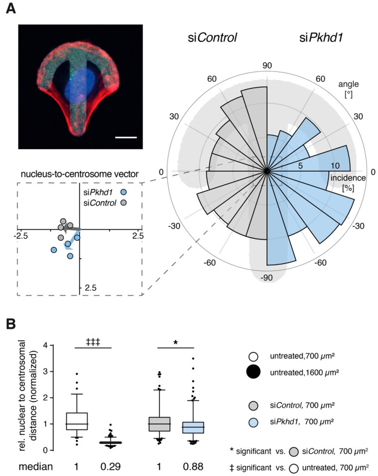
Figure 3.
Defective positioning of centrosomes in 1 and 2-cell stages. (A) Adhesion of single cells on crossbow-shaped micropatterns (upper left) enforces positioning of centrosomes, in front of nucleus, corresponding to the polarized orientation of a migratory cell [22] (F-actin (red), nucleus (blue), centrosomes (green), pattern (white)). For siControl and siPkhd1-treated cells (right), angle distributions of nucleus-to-centrosome vectors are shown to mirror symmetrically (−90° to 90°). Mean vectors of independent experiments (lower left), as calculated from the total range (−180° to 180°), and total mean vector reveal a defect of siPkhd1 cells in their ability to correctly place centrosomes towards the migratory front. (n =250–310 cells, n = 4 independent experiments; p = 0.0026; non-parametric test for circular data corresponding to 1-way ANOVA [23]). Size bar, 10 µm. (B) In the 2-cell stage, central positioning of centrosomes is critical to initiation of lumen formation and is suppressed by strong peripheral contractility [13]. Using 2-cell stages, relation of relative nuclear to centrosomal distances was determined in untreated cells, on 700 and 1600 µm2, and in siPkhd1 and siControl-treated cells on 700 µm2. Graphical illustration is provided in Supplementary Figure S4D. (n = 2/5 independent experiments, >130/180 cell pairs per condition for untreated and siRNA-treated cells, respectively; non-parametric t-test/Mann–Whitney, p < 0.001/0.05, ‡‡‡/*).

