Figure 1.

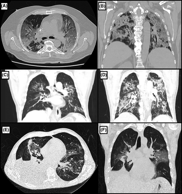
A and B, Thoracic computed tomography (CT) scan of patient 2, showing bilateral several ground‐glass pulmonary opacities affecting approximately 50% of the lungs, occasionally associated with thickening of interlobular septa and thin reticulate, in addition to peripheral sparse consolidation foci with greater extension in the posterior aspect of the lower lobes (A—axial view, B—coronal view). C, Thoracic CT scan of patient 3 on 10th postoperative day (POD), showing bilateral multiple ground‐glass pulmonary opacities, sometimes associated with thickening of interlobular septa and fine reticulate, affecting less than 50% of the lungs. D, Thoracic CT scan of the same patient on 20th POD, performed due to shortness of breath worsening, revealing increase in number and dimensions of ground‐glass pulmonary opacities, now affecting more than 50% of the lungs. E and F, Thoracic CT scan of patient 5, showing numerous bilateral peribronchovascular ground‐glass opacities, mainly in the upper lobes, some with thickening of the inter and intralobular septa. There is also a large pleural effusion on the right side with restrictive atelectasis of the adjacent pulmonary parenchyma (E—axial view, F—coronal view)
