Fig. 7.


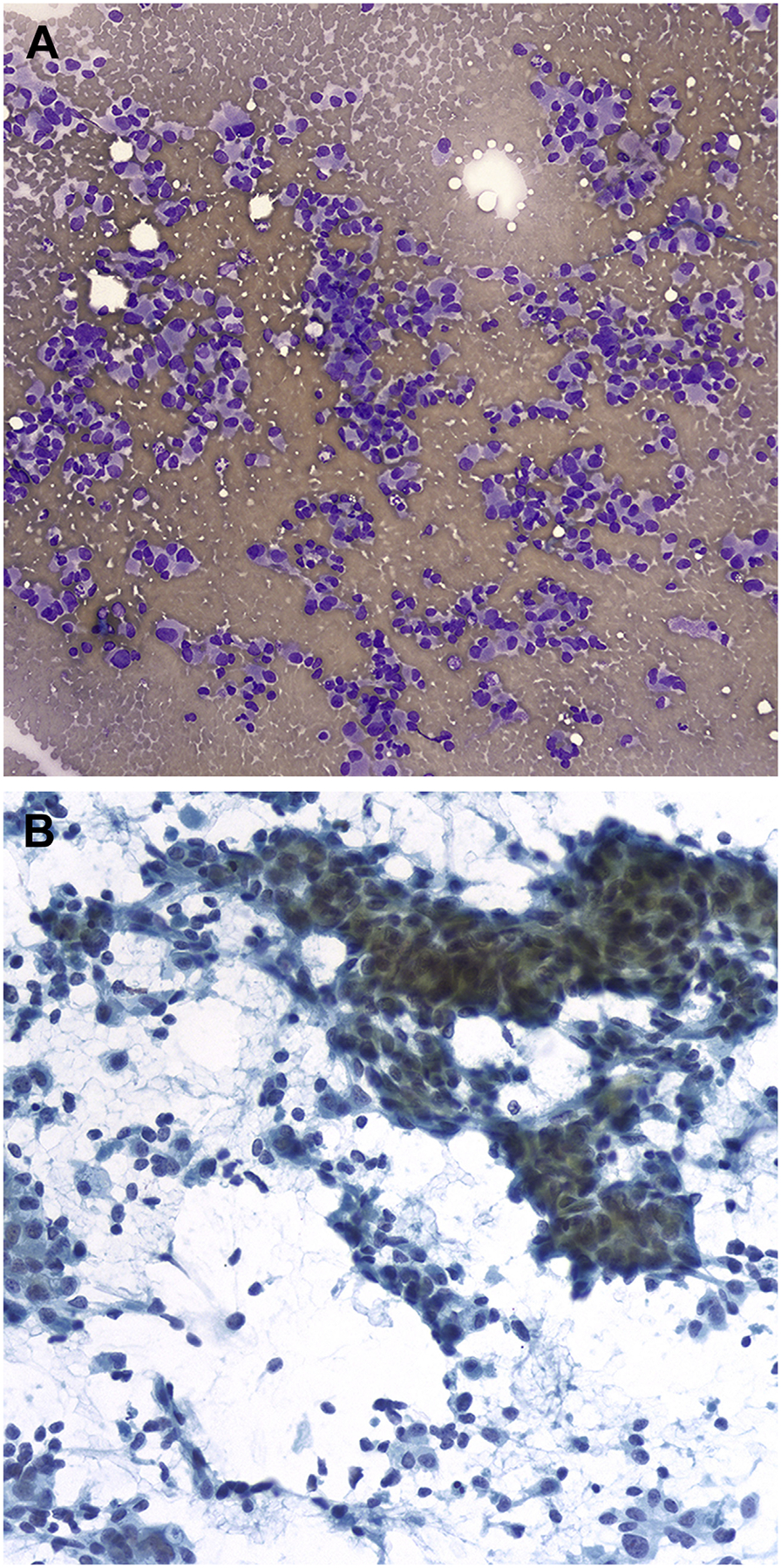
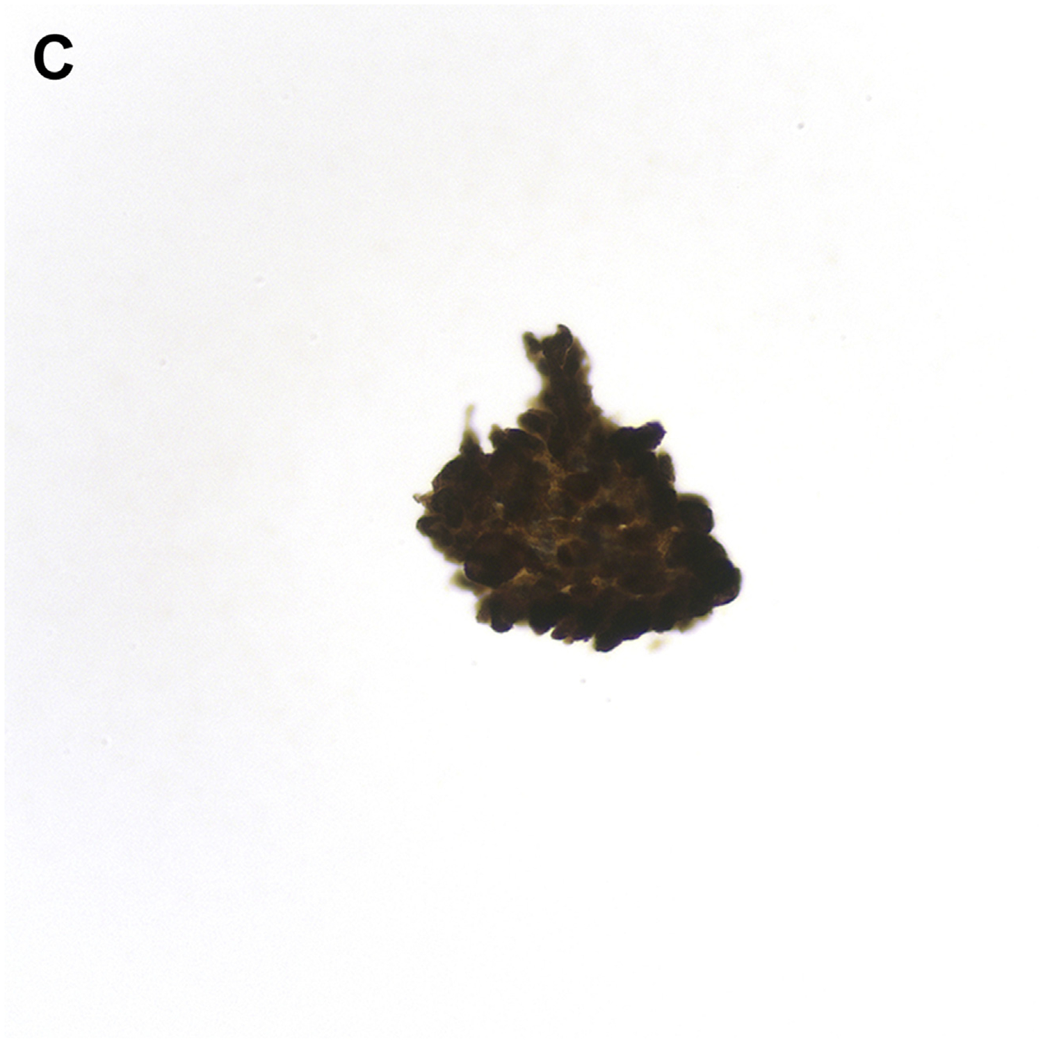
FNA of MTC. (A) Loose clusters and single cells with eccentrically placed nuclei (Diff-Quik stain, original magnification ×200). (B) The tumor shows spindling with a pseudopapillary appearance, nuclear elongation, and rare grooves. Single cells are also present in the background (Papanicolaou stain, original magnification ×400). (C) Tumor cells are immunochemically positive for calcitonin. (Avidin-Biotin complex method, original magnification ×400).
