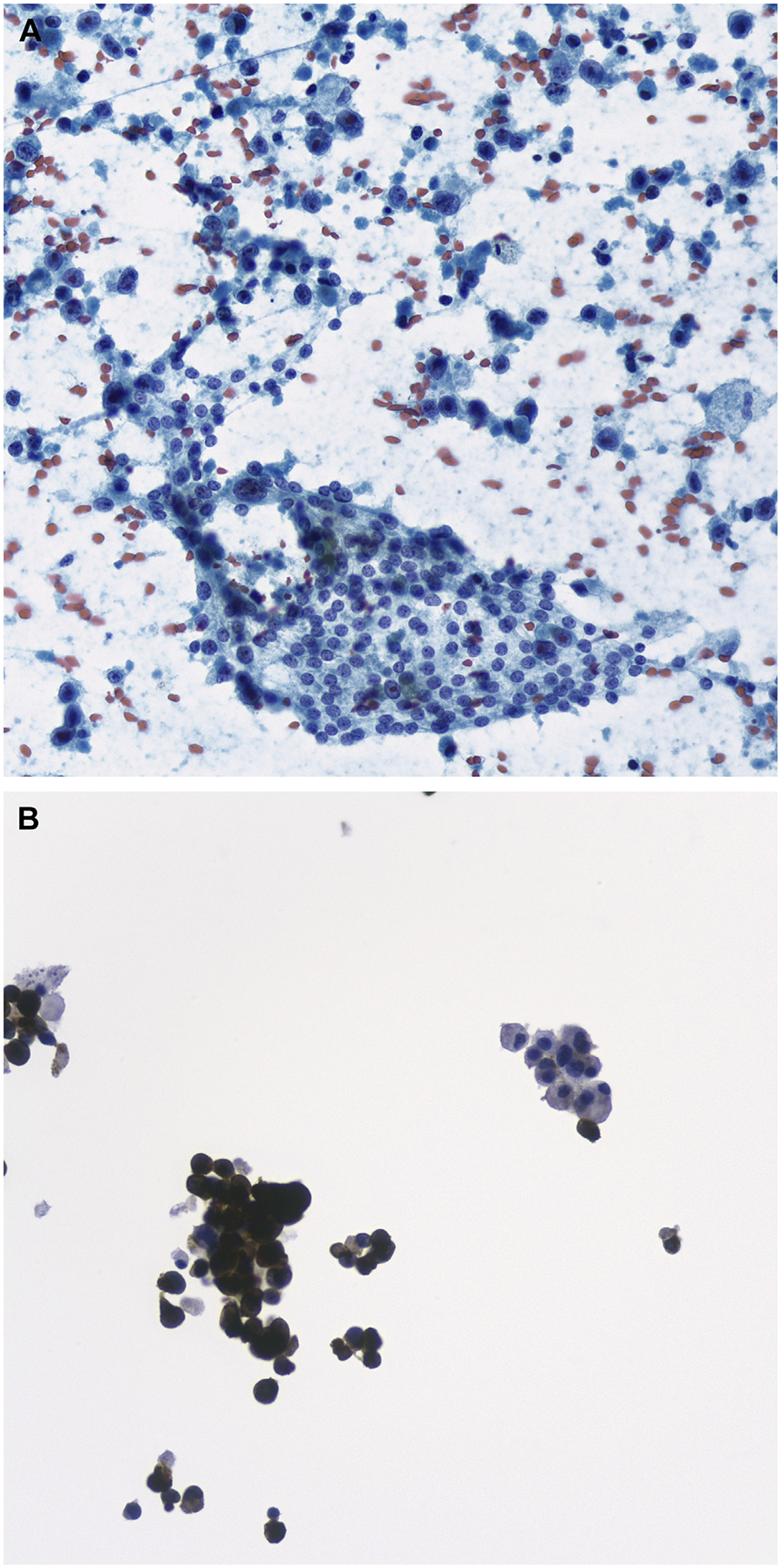
Fig. 9.

FNA of metastatic melanoma to the thyroid. (A) Infiltrating single cells of melanoma are seen in the background of nonneoplastic follicular cells (Papanicolaou stain, original magnification ×400). (B) Melan A immunochemical stain highlighting the tumor cells; the background normal follicular cells are negative. (Original magnification ×400).
