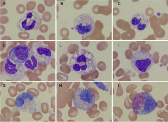
FIGURE 1.

Images of myeloid forms seen in children with multisystem inflammatory syndrome (MIS‐C). A‐C, Images from patient A. A, Neutrophil with toxic granulation and heavy vacuolization. B, An early myeloid form with vacuolization. C, A neutrophil with presence of Dohle bodies. D‐F, Images from patient B. D, An early myelocyte, metamyelocyte, and neutrophils with vacuolization and toxic granulation. E, A neutrophil with significant vacuolization and granulation. F, An early myeloid form, likely metamyelocyte. G‐I, Images are from patient C. G, A late metamyelocyte with vacuolization. H, An early metamyelocyte. I, An eosinophilic myelocyte. Together these images suggest significant inflammation with vacuolated myeloid forms and the presence of toxic granulation. The early myeloid forms (bands, metamyelocytes, and myelocytes) are seen in high number in the peripheral smears of children with MIS‐C
