Figure 1.
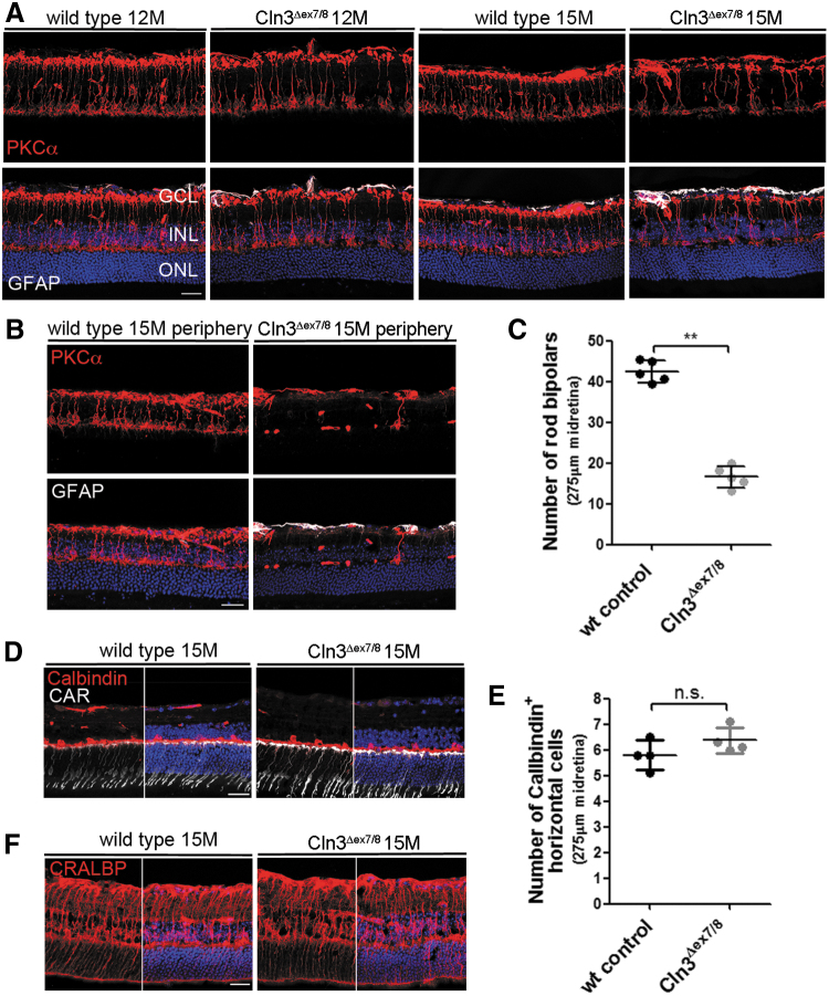
Cln3Δex7/8 mice show a progressive loss of rod bipolar cells. (A) Representative confocal images of the midretina from Cln3Δex7/8 and wild-type mice immunostained with antibodies against PKCα and GFAP. At 12 months, fewer PKCα-positive cells were detected in mutant than wild-type retinas, which progressed to an obvious loss of cells at 15 months. GFAP staining did not appear to be elevated. (B) PKCα staining revealed a pronounced lack of rod bipolar cells in the periphery of the retina. (C) Quantification of the number of PKCα-positive rod bipolar cells in the midretina of mutant and wild-type retinas at 15 months. Wild-type and Cln3Δex7/8: n = 5 eyes. Data are presented as mean ± SD and analyzed by a nonparametric Mann–Whitney test (**p = 0.0079). (D, F) Representative images of 15-month-old mutant and wild-type midretina immunostained for Calbindin, CAR, and CRLABP. No obvious loss of horizontal, cone-photoreceptor, and Mueller glial cells was observed. (E) Quantification of the number of Calbindin-positive cells in the INL did not reveal any significant difference between mutant and wild-type retinas at 15 months. Wild-type and Cln3Δex7/8: n = 4 eyes. Data are presented as mean ± SD. Scale bar: 25 μm. CAR, cone-arrestin; CRLABP, cellular retinaldehyde binding protein; GCL, ganglion cell layer; GFAP, glial fibrillary acidic protein; INL, inner nuclear layer; ONL, outer nuclear layer; PKCα, protein kinase C alpha-subunit; SD, standard deviation.

