Figure 4.
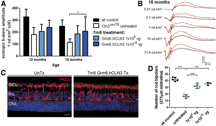
(A) Scotopic ERG b-wave amplitudes of 7m8.Grm6.hCLN3-treated mutant mice. Wild type: n = 7–8 eyes, Cln3Δex7/8: n = 8–9 eyes, 7m8.CMV.hCLN3 1 × 10⁹ vg: n = 5–6 eyes, 7m8.CMV.hCLN3 1 × 10¹⁰ vg: 3–4 eyes. Wild-type recordings from Fig. 1 were added as a reference. (B) Scotopic ERG traces of a mutant mouse that received treatment in one eye (red). The contralateral eye was left uninjected (black). (C) Representative confocal images of the midretina from untreated and a high-titer 7m8.Grm6.hCLN3-treated eyes stained for PKCα at 15 months. (D) Counts of PKCα-positive cells in the midretina at 15 months. Data are presented as mean ± SD and analyzed by (A) two-way ANOVA with Bonferroni test and (D) one-way ANOVA with Kruskal–Wallis test (*p < 0.05, ***p < 0.001). Scale bar: 25 μm.

