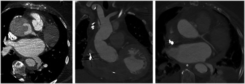
Figure 3.

Pre‐ and postoperative computed‐tomography scans of patient 2. Figures show axial view of the dissected aortic root and descending aorta (left), postoperative coronal scan of the ascending aorta (middle), and an axial view of postoperative aortic root and descending aorta (right)
