Figure 5.
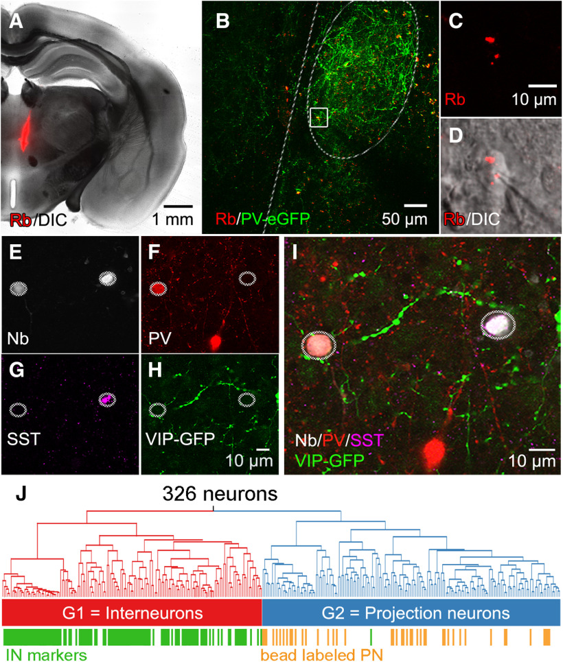
Identification of claustrum PNs and INs. Retrobead (Rb) labeling was used to identify PN, while marker protein expression was used to identify IN. A, Overview of a Rb injection (pseudocolored red) targeting ventral and medial thalamic nuclei. B, Coronal brain slice from the same animal in A. The claustrum (dashed outline) can be identified by strong PV-eGFP expression (green), while subcortical-projecting neurons are labeled in red. C, High-magnification image of Rb-label (pseudocolored red) in the subcortical-projecting claustrum neuron shown within the white square in panel B. D, Combined Rb and DIC image showing the same Rb-labeled neuron and the glass recording electrode. E, Ovals indicate two neurobiotin-labeled claustral neurons. The left neuron expressed PV (F), while the right neuron expressed SST (G), and neither neuron expressed VIP-promoter-driven eGFP (H). I, Merger of E–H indicates that the left cell is a PV-IN, while the cell on the right is an SST-IN. J, Dendrogram from Figure 3, annotated with identified PN that were labeled with Rb (orange) and IN that expressed PV, SST, or VIP (green). IN markers were almost exclusively restricted to cells in the left arm of the dendrogram, identifying G1 as IN, while retrobead-labeled neurons were exclusively in the right arm of the dendrogram containing virtually no neurons expressing IN markers, indicating that G2 cells are PN.

