Figure 4.
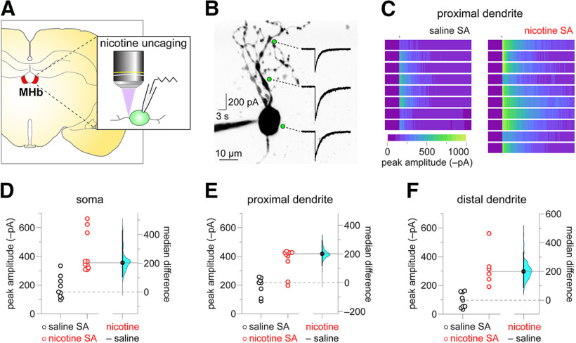
Nicotine SA boosts nAChR function at the soma and in dendrites. A, Experiment schematic. Coronal brain slices containing MHb were prepared from rats after the last SA session. MHb neurons were patch clamped, neuronal morphology was imaged with 2PLSM, and nicotine was uncaged via laser flash photolysis. B, Representative nicotine uncaging experiment. An exemplar two-photon image of a MHb neuron is shown, along with locations (soma, proximal dendrite, distal dendrite) where nicotine was uncaged and the recorded nAChR response to such uncaging. C, Proximal dendrite uncaging responses. Heat map representations of proximal dendrite nicotine uncaging responses from individual neurons are shown for nicotine SA and saline SA rats. All heat maps are scaled to the response with the greatest magnitude (a nicotine SA response). D–F, Gardner–Altman plots of peak nicotine uncaging currents at the soma (D), proximal dendrite (E), and distal dendrite (F). At left, scatter plots show individual uncaging currents for nicotine SA and saline SA rats. At right, the effect size (median difference) and bootstrap 95% CI are shown.

