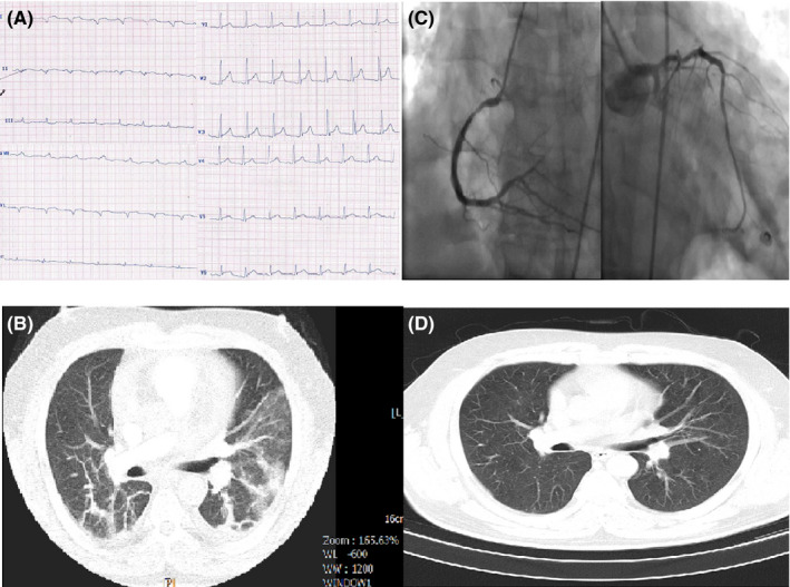
FIGURE 1.

A, ECG of case 1 showing a nonspecific pattern: normal sinus rhythm (NSR), right axis deviation, left atrial abnormality, widening of QRS in limb leads and tall R wave in V1‐V3 as well as ST elevation in V2‐V6. B, Chest CT of case 1 showing typical ground‐glass opacification in both lungs. C, Selective coronary angiography of case 1 revealing three‐vessel disease. D, Chest CT of case 1 after treatment without coronavirus involvement
