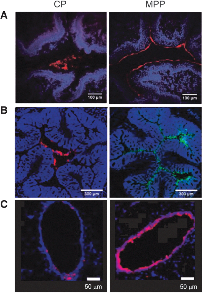
FIG. 4.

Distributions of MPPs and CPs in the mucus layer of various organs of the mouse model (obtained by fluorescent imaging of tissue cross-sections collected after administration of CP and MPP suspensions; red or green represents fluorescence from the nanoparticles; blue represents fluorescence from the stained tissue). (A) MPPs (particle diameter ∼110 nm) and CPs (particle diameter ∼90 nm) in mouse vagina. Reprinted from Ensign et al., Copyright © 2012, with permission from American Association for the Advancement of Science10. (B) MPPs and CPs (both ∼100 nm in diameter) in mouse colorectum. Reprinted from Maisel et al., Copyright 2015, with permission from Elsevier11. (C) MPPs (particle diameter ∼60 nm) and CPs (particle diameter ∼50 nm) in mouse large airway. Reprinted from Suk et al., Copyright 2014, with permission from Elsevier.12
