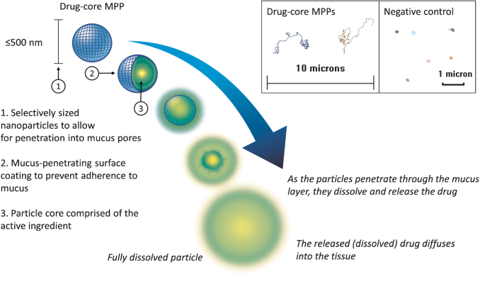
FIG. 7.
Schematic depiction of a drug-core MPP. The inset is showing 15-s trajectories of drug-core MPPs (particle size of ∼270 nm in this example) and a negative control (200 nm polystyrene nanoparticles) in human cervicovaginal mucus as observed by fluorescence microscopy. The inset is adapted from Cu et al.61

