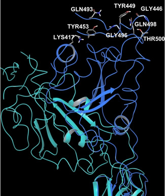
Figure 1.

SARS‐CoV‐2 PDB structures superposition unveiling RBD dynamic behaviour. On the left, overlap of SARS‐CoV‐2 S trimers in closed (PDB 6VXX) and open state (PDB 6VYB); on the right, a close‐up: the light blue structure shows PDB 6VXX S protein, while the blue chain exhibits the open state of PDB 6VYB S protein. PDB 6VXX: green, violet and light blue chains in closed conformations; PDB 6VYB: yellow, pink and blue chains in open conformations.
