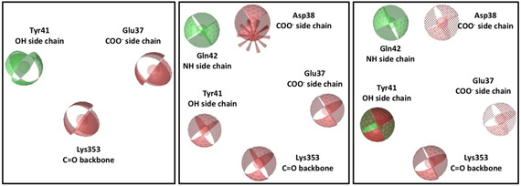
Figure 4.

Pharmacophore maps built on RBD N‐terminal region. On the left, 6M17 Pharmacophore map; in the middle, 6M0J Pharmacophore map; on the right, shared pharmacophore map. Red spheres are hydrogen‐bond acceptors, green spheres are hydrogen bond donors, green‐red sphere is both hydrogen‐bond donor and acceptor, red spike is a negative ionisable feature and dotted spheres are features marked as optional.
