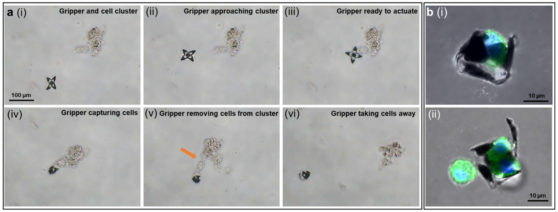
Figure 5. Few-cell biopsy from a cell cluster and visualization of cellular components of captured suspended cells.

(a) The process of a gripper capturing and excising cells from a cell cluster in the following steps, (i) a gripper and a cell cluster at a distance; (ii) the gripper approached the cell cluster guided by magnetic field; (iii) the gripper reached the desired location; (iv) upon temperature increase, the gripper grasped a few cells; (v) the gripper was dragging the captured cells away from the cluster by changing directions of magnetic field; (vi) the gripper successfully excised cells and moved them away. (b) Immunofluorescence images of suspended fibroblast cells captured by the grippers: (i) side view of a captured single cell, (ii) two cells captured inside a gripper. Cells were fixed and stained for nuclei (DAPI, blue), and β-tubulin (antibody labeling, green).
