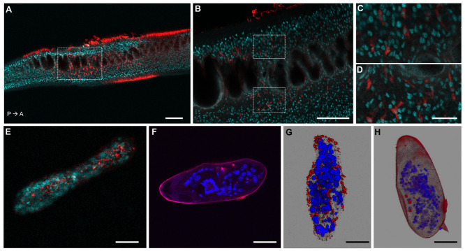
Figure 2. Confocal microscopy images of S. mansoni parasites transfected with fluorescently labelled Cas9-gRNA (ATTO™ 550 signal in red), fixed and DAPI-stained (DAPI signal in aqua blue or blue).
( A) Confocal optical section of a male adult worm. P→ A: indicates the posterior anterior axis. Scale bar: 100 µm. ( B) Magnified squared-area in ( A). Scale bar: 50 µm. ( C, D) Magnified top and bottom squared-areas in ( B), respectively. Scale bar: 10 µm. ( E, F) Confocal optical sections of a sporocyst and an egg, respectively. ( G, H) Maximum intensity projection of z-stack images of a sporocyst and an egg, respectively. Scale bars in E– H: 25 µm. The images of worms, sporocysts and eggs, were taken from representative specimens collected from the biological replicate "Experiment 7, tags 50 and 61", "Experiment 11, tag 19 (panel E) and "Experiment 2, tag 6 (panel G)", and "Experiment 1, tag 4", respectively.

