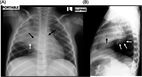
Figure 1.

A, A supine frontal chest radiograph was performed as a portable examination. There is confluent air‐space disease in the right upper and middle lobes with a small focus of breakdown (white arrow) and airway narrowing at the left main bronchus and the bronchus intermedius (black arrows), which represent surrogate markers of hilar and mediastinal lymphadenopathy. In conjunction with the air‐space disease, the parenchymal breakdown and airway narrowing support a diagnosis of primary pulmonary tuberculosis, which was confirmed using Gene Xpert on a gastric washing sample. B, Lateral radiograph confirms the area of parenchymal breakdown (black arrow), presumably in the right middle lobe, and also confirms the lymphadenopathy (white arrows) posteroinferiorly to the bronchus intermedius
