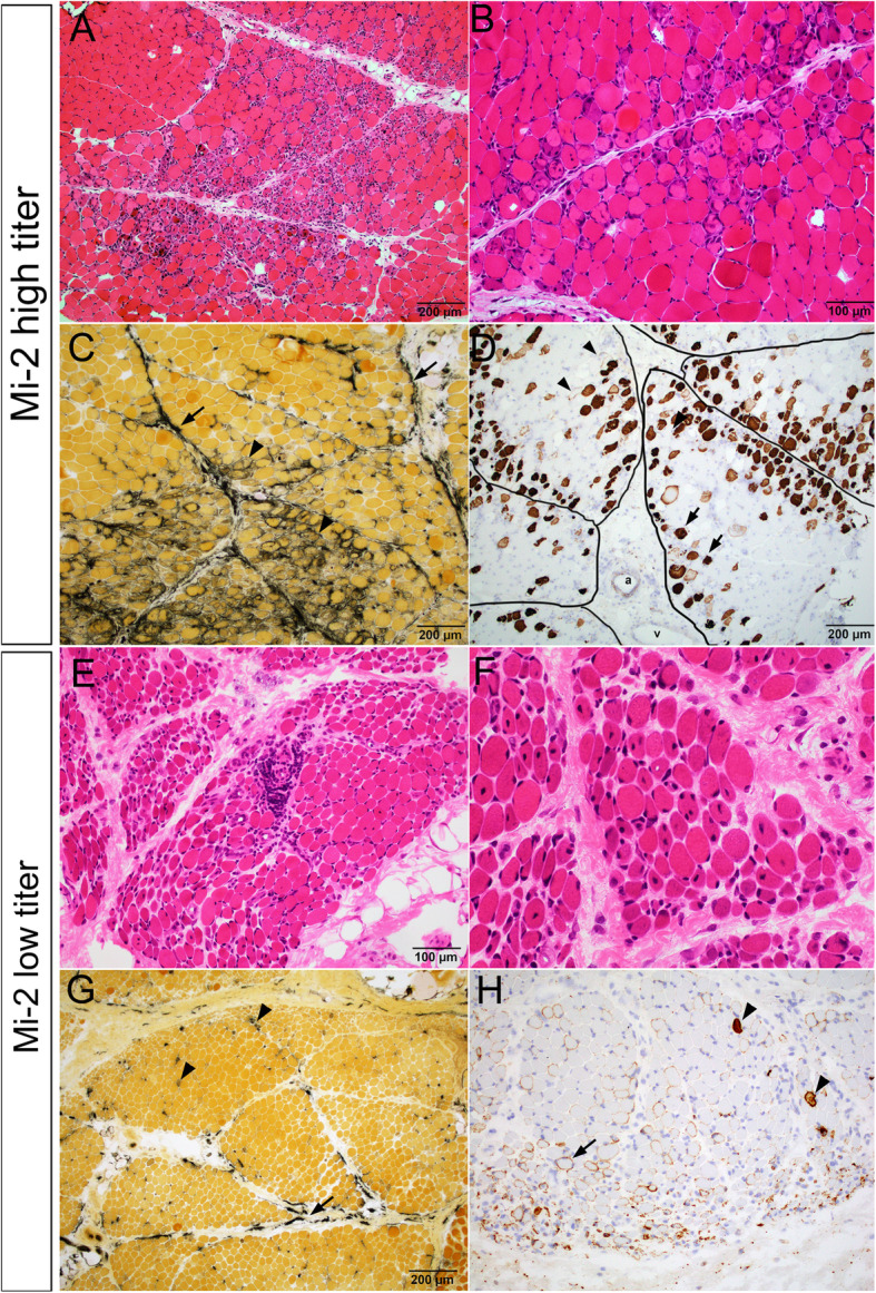
Fig. 2.

Characteristic muscle pathology in patients with high and low titers of Mi-2 auto-antibody. a-d Patient 4 with high Mi-2 titer. a Low and (b) high power images of H&E stained cryosections showed prominent perifascicular myofiber necrosis and regeneration. c Alkaline phosphatase stain highlighted frequent regenerating fibers (arrow heads) as well as strong perimysial connective tissue reactivity (arrows). d C5b-9 immunostain showed frequent necrotic fibers concentrated in the perifascicular regions (arrow heads) but might present at the center of fascicles. Viable but injured myofibers might show sarcolemmal C5b-9 reactivity (arrow heads). There was no significant capillary C5b-9 reactivity. Perimysium was outlined by black lines. “a” indicate perimysial artery. “v” indicate perimysial vein. e-h Patient 8 with low Mi-2 titer. e Low and (f) high power H&E showed prominent perifascicular atrophy and frequent internal nuclei, but few acutely necrotic fibers. g Alkaline phosphatase stain highlighted rare regenerating fibers (arrow heads) and patchy discontinuous perimysial connective tissue reactivity (arrows). h C5b-9 immunostain highlighted occasional necrotic fibers (arrow heads) but rather wide spread sarcolemmal C5b-9 reactivity in viable myofibers (arrow heads). There was no significant capillary C5b-9 reactivity
