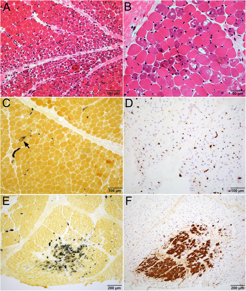
Fig. 3.

Characteristic muscle pathology in patients with high titers of NXP-2 auto-antibody. a-f Patient 9 with high NXP-2 titer. a Low power image of H&E stained cryosections showed perifascicular atrophy. High power image (b) showed that many myofibers had basophilic vacuolar degeneration. c Alkaline phosphatase stain highlighted only rare regenerating fiber; the basophilic vacuolar fibers were non-reactive. There was no abnormal connective tissue reactivity. d C5b-9 immunostain highlighted prominent capillary reactivity, but no necrotic fibers or sarcolemmal C5b-9 reactivity. Panels (e) alkaline phosphatase and (f) C5b-9 showed a focus of infarction with grouped necrotic and regenerating fibers at the center of a fascicle
