
FIGURE 1.
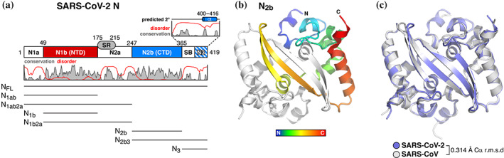
Structure of the severe acute respiratory syndrome coronavirus 2 (SARS‐CoV‐2) nucleocapsid dimerization domain. (a) Domain structure of the SARS‐CoV‐2 nucleocapsid protein, as defined previously, 46 , 47 with plot showing the Jalview alignment conservation score (three‐point smoothed; gray) 61 and DISOPRED3 disorder propensity (red) 62 for nine related coronavirus N proteins (SARS‐CoV, SARS‐CoV‐2, Middle‐East respiratory syndrome [MERS]‐CoV, HCoV‐OC43, HCoV‐HKU1, HCoV‐NL63, and HCoV‐229E, infectious bronchitis virus [IBV], and Murine Hepatitis virus [MHV]). SR, serine/arginine rich domain; SB, spacer B. The boundary between SB and N3 is not well defined due to low identity between SARS‐CoV/SARS‐CoV‐2 and MHV N proteins. 47 All purified truncations are noted at bottom. (b) Top‐down view of the SARS‐CoV‐2 N2b dimer, with one monomer colored as a rainbow (N‐terminus blue, C‐terminus red) and the other colored white. See Figure S1a for comparison with other structures of this domain. (c) Structural overlay of the SARS‐CoV‐2 N2b dimer (blue) and the equivalent domain of SARS‐CoV‐N (PDB ID 2CJR) 34