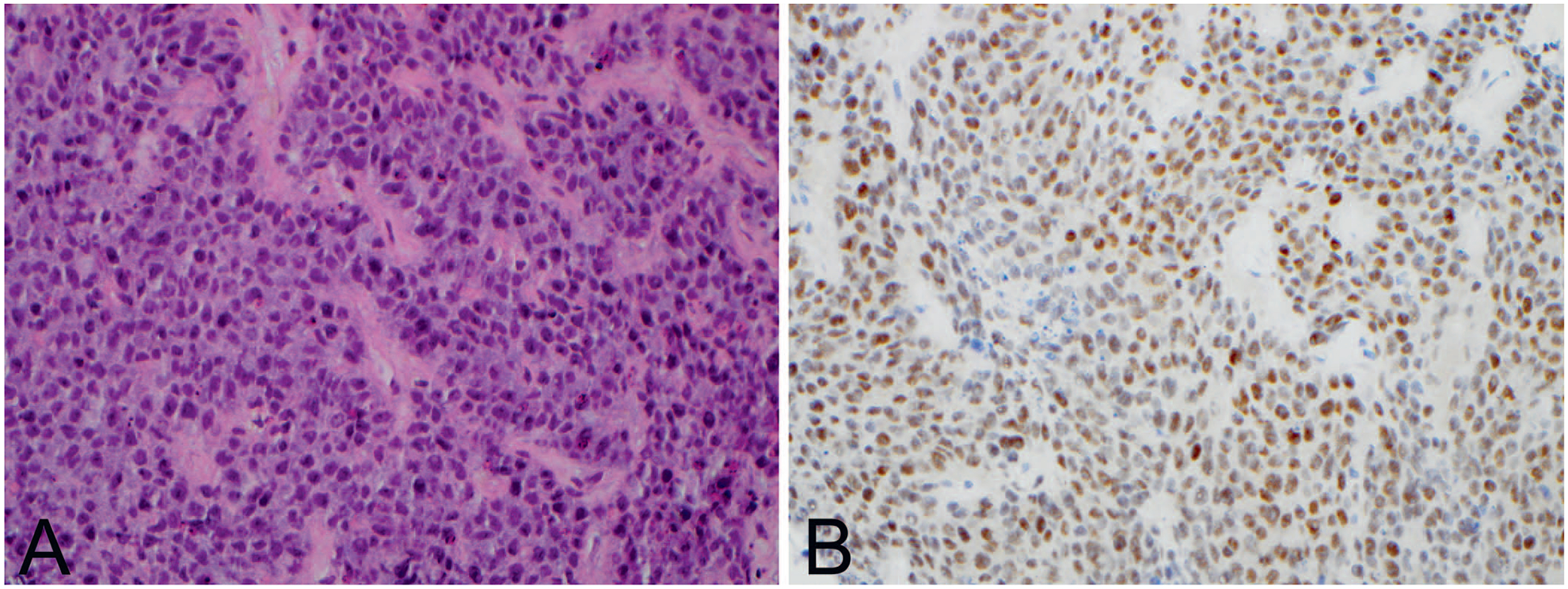
Figure 1.

NUT midline carcinoma. A, Tumor is composed of sheets of monomorphic poorly differentiated cells with scattered apoptotic bodies. B, Immunohistochemistry reveals positive nuclear expression with NUT antibody (hematoxylin-eosin, original magnification ×40 [A]; original magnification ×40 [B]).
