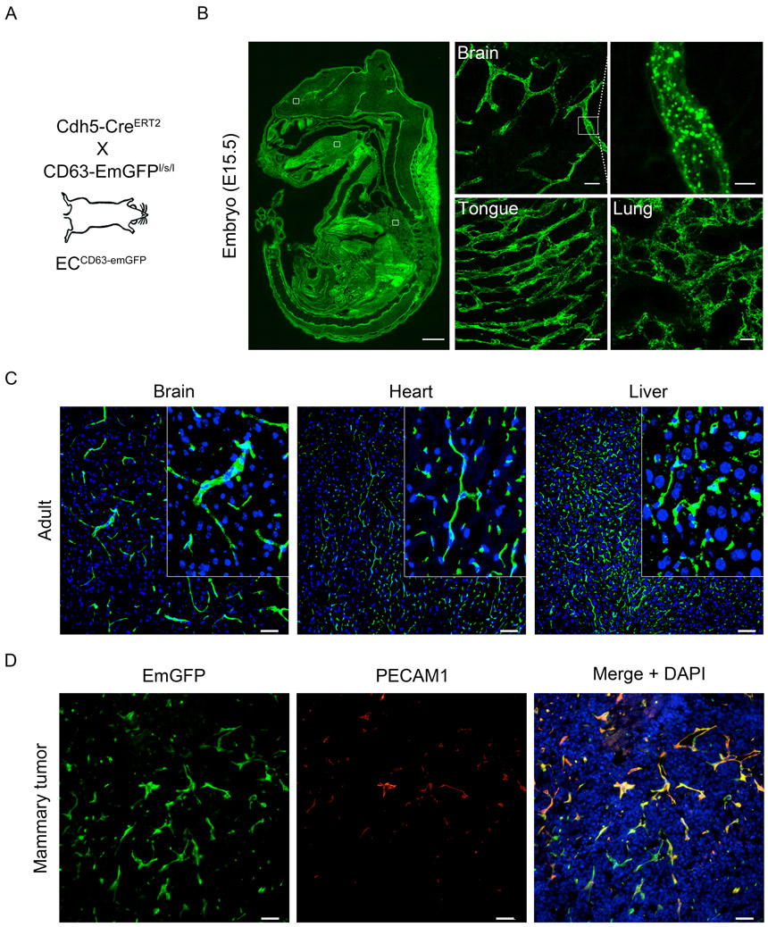
Fig 3. The vasculature in Cdh5-CreERT2:CD63-emGFPl/s/l mice is labeled with GFP.
(A) Schematic summarizing the mouse cross. (B) Blood vessels are labeled in pregnant mice that receive tamoxifen to induce emGFP expression in the developing vasculature of embryonic tissues are organs. The inset at right of the brain section shows emGFP+ puncta that are likely CD63+ endosomes. Scale bar: whole embryo = 1mm, organs = 25 μm, and enlarged brain vessels = 5 μm. (C) Native emGFP fluorescence using fresh cryosections from brain, heart, and liver. The inset shows clear vascular expression. Nuclei are labeled with DAPI. (D) Angiogenic vasculature in EO771 mammary tumors is also emGFP+ and co-localizes with the pan EC marker PECAM1. (D)

