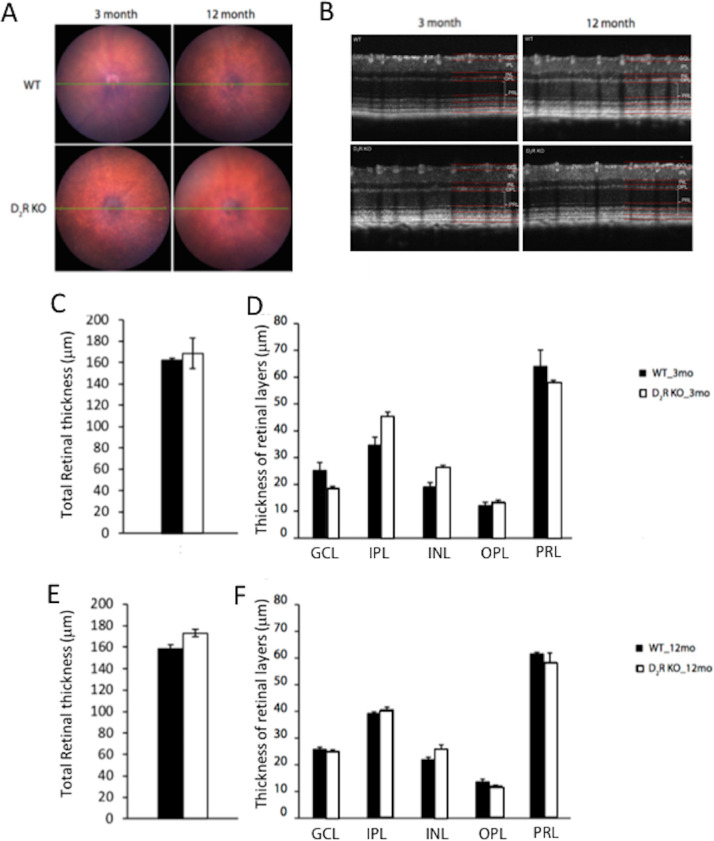
Figure 6.
Retinal thickness is unaltered by the lack of D2R signaling. (A) Fundus images acquired from WT and D2R KO mice of 3 and 12 months of age did not show any significant difference between the two genotypes at both ages. (B) Total retinal thickness was not different between the two genotypes at both the age groups. (C, E). No differences were detected in the thickness of the different retinal layers between the two genotypes at both ages (D, F). We used t-tests, P > 0.05 in all cases. Bars represent the means ± SEM (N = 4–6). GCL, ganglion cells layer, INL, inner nuclear layer; IPL, inner plexiform layer; OPL, outer plexiform; PRL, photoreceptor layer).

