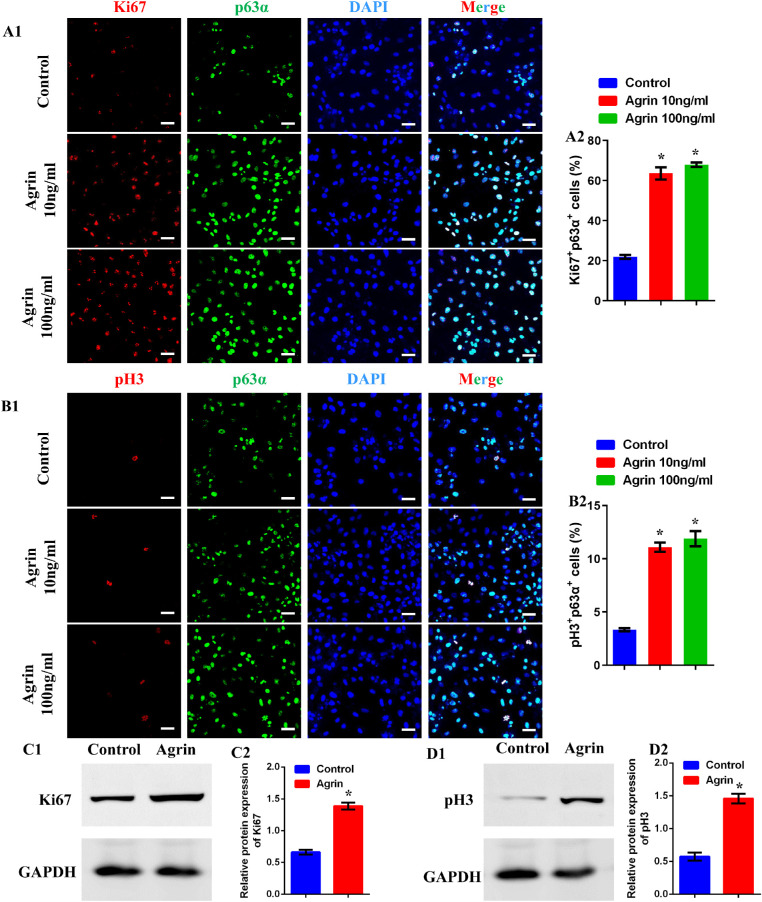
Figure 2.
Agrin promoted the proliferation of limbal stem cells in vitro. (A1) Representative images of cultured limbal stem cells stained with DAPI (blue), p63α (green), and Ki67 (red) after treatment with Agrin (0, 10, and 100 ng/mL) for 2 days, scale bar = 20 µm. (A2) Percent of proliferating limbal stem cells (p63α+ Ki67+) in response to 0, 10, and 100 ng/mL Agrin for 2 days, n = 1014, 2696, and 2654 limbal stem cells pooled from five samples (eight microscopic fields for one sample) in 0, 10, and 100 ng/mL Agrin groups. * P < 0.05 vs. control. (B1) Representative images of cultured limbal stem cells stained with DAPI (blue), p63α (green), and pH3 (red) after treatment with Agrin (0, 10, and 100 ng/mL) for 2 days, scale bar = 20 µm. (B2) Percent of proliferating limbal stem cells (p63α+ pH3+) in response to 0, 10, and 100 ng/mL Agrin for 2 days, n = 1022, 2613, and 2640 limbal stem cells pooled from five samples (eight microscopic fields for one sample) in 0, 10, and 100 ng/mL Agrin groups. * P < 0.05 vs. control. (C) Representative images (C1) and quantification (C2) of Ki67 protein expression in limbal stem cells analyzed by Western blotting with or without 10 ng/mL Agrin for 2 days. GAPDH was used as control. n = 6 samples for each group. * P < 0.05 vs. control. (D) Representative images (D1) and quantification (D2) of pH3 protein expression in limbal stem cells analyzed by Western blotting with or without 10 ng/mL Agrin for 2 days. GAPDH was used as control. n = 6 samples for each group. * P < 0.05 vs. control.

