Figure 5.
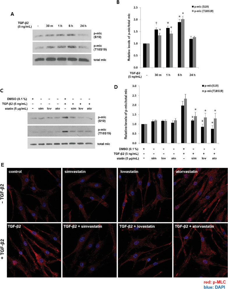
Statins suppress TGF-β2-induced phosphorylation of MLC in astrocytes from human ONH. Serum-starved astrocytes were incubated with TGF-β2 (5 ng/mL) for the indicated periods without (A, B) or after (C, D) prior incubation with simvastatin, lovastatin, and atorvastatin (5 µg/mL) for 1 hour followed by TGF-β2 (5 ng/mL) for 6 hours. Amounts of pMLC at S19 and at T18S19 were determined by western blotting and are presented as fold changes relative to untreated (B) or DMSO (D). Compared with untreated cells, MLC phosphorylation was induced within 30 minutes and peaked at 6 hours (A, B, *P < 0.001, †P < 0.05 vs. untreated; n = 3). Effects of statins on pMLC were compared between cells incubated with or without statins, followed by TGF-β2. Statins suppressed TGF-β2-mediated phosphorylation of MLC (D, *P < 0.05 vs. TGF-β2 + DMSO; n = 3). (E) Cells were incubated with TGF-β2 (5 ng/mL) without or with prior incubation with statins (5 µg/mL) for 1 hour, then stained with pMLC (S19) antibody. Cell nuclei were counterstained with DAPI. Specific cytoplasmic immunoreactivity for pMLC was increased by TGF-β2 and suppressed by statins.

