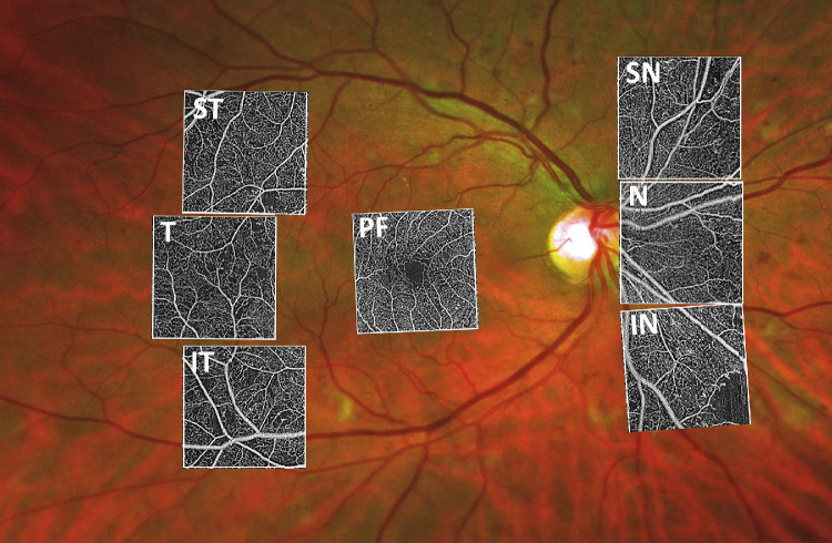
Figure 1.
The 15 × 15 mm OCTA montage (Plex Elite, Zeiss) of a patient with severe diabetic retinopathy. The position of the seven 3 × 3 mm areas imaged according to our scanning protocol are outlined in the white boxes. PF = parafovea, T = temporal, ST = superior temporal, IT = inferior temporal, N = nasal, SN = superior nasal, IN = inferior nasal.

