


Figure 1.
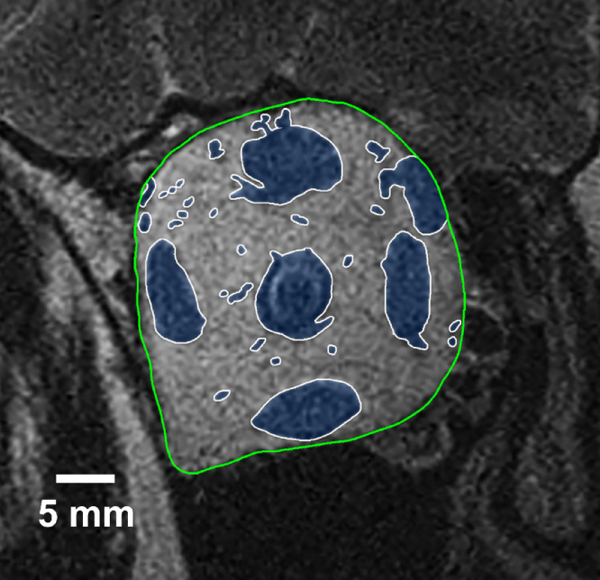
Quasicoronal MRI in the mid-portion of a representative right orbit, whose bony walls are outlined in green. Intraorbital structures including extraocular muscles, ON, and blood vessels, are outlined in white and shaded blue. Orbital fat represents the remaining orbital area excluding structures shaded in blue.