


Figure 2.
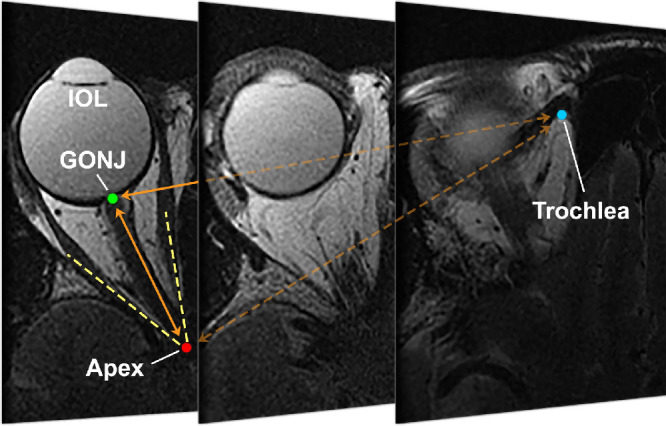
Axial MRI of representative orbit in pseudophakic subject, with contiguous 2 mm thick image planes arranged from inferior at mid-globe at left, to superior at right with the plane including the trochlea. GONJ, globe-optic nerve junction; IOL, intraocular lens.