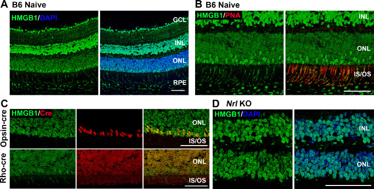
Figure 1.
The expression pattern of HMGB1 in the mouse retina. (A) Naive retinas of 8-week-old C57BL/6J mice were probed with anti-HMGB1 antibody (Ab). Note low HMGB1 Ab IF in most of the nuclei in the ONL. (B) Naive retinas of C57BL/6 mice were probed with anti-HMGB1 Ab (green) and PNA (red). Note juxtaposition of PNA binding (indicating cone inner and outer segments) with a minority of photoreceptor nuclei with high HMGB1 Ab binding. (C) Colocalization of HMGB1 (green) and Cre (red) in the naive retinas of opsin-Cre and rhodopsin-Cre (Rho-cre) mice on a C57BL/6J background. Note the colocalization of high HMGB1 IF with opsin-Cre IF but no colocalization with rho-Cre IF. (D) Naive retinas of Nrl−/− mice (cone-like photoreceptors only) were probed with anti-HMGB1 Ab and nuclei were counterstained with DAPI. Note that HMGB1 IF is comparable in the ONL and INL. GCL, ganglion cell layer; INL, inner nuclear layer; IS/OS, inner and outer segments. Scale bars: 50 µm.

