Figure 2.
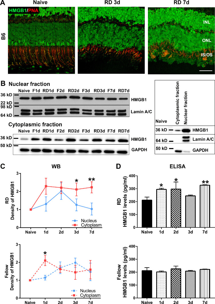
RD triggers the translocation and release of HMGB1. RD was created by subretinal injection of 1% hyaluronic acid in the left eyes of C57BL/6 mice. The contralateral eyes did not receive any treatment and served as fellow controls. (A) Costaining of HMGB1 Ab (green) and PNA (red) in the naive retina and detached retinas at 3 and 7 dprd. Note the relatively low HMGB1 IF in most PR nuclei and the spatial juxtaposition of high HMGB1 IF in some PR nuclei with PNA staining of cone inner and outer segments. Scale bar: 50 µm. (B–D) Mouse eyes were enucleated at 1, 2, 3, and 7 dprd. Eyecups were minced and supernatants collected for ELISA to measure the levels of soluble HMGB1. (B) Insoluble eyecup pieces underwent extraction of nuclear and cytoplasmic proteins for Western blot. Lamin A/C and glyceraldehyde 3-phosphate dehydrogenase were used as loading controls for the nuclear fraction and cytoplasmic fraction, respectively. Blots on the right side show the relative amounts of HMGB1 protein in cytoplasmic and nuclear fractions in naive retinas and the efficiency of fractionization. (C) Graphs show the quantification of protein levels based on densitometry of the Western blots (n = 3). (D) Graphs show levels of HMGB1 in the eyecup supernatant (n = 4) measured by ELISA. Data were plotted with mean ± SD. One-way ANOVA. *Indicates significant difference in comparison with naive. Statistical significance was set at P < 0.05. F, fellow.

