Figure 3.
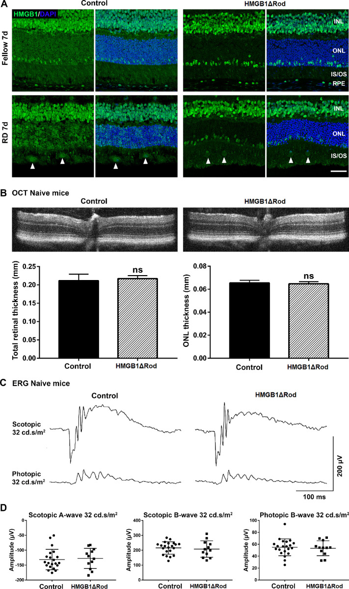
Conditional knockout of HMGB1 in rod photoreceptors does not affect retinal thickness or ERG response at baseline. (A) Fellow and detached retinas at 7 dprd were probed with HMGB1 Ab to confirm the complete and specific knockout of HMGB1 in rod photoreceptors in HMGB1ΔRod mice. Rho-Cre+ mice served as controls. Nuclei were counterstained with DAPI. Arrowheads show the infiltrated cells positive for HMGB1 IF. Scale bar: 50 µm. (B–D) OCT imaging and ERG analyses were performed in naive HMGB1ΔRod and control mice at 20 weeks of age. Represented OCT images are shown in (B). The thicknesses of the total retina and ONL were measured at four points (nasal, temporal, superior, and inferior) at a distance of 500 µm from the optic nerve head. Represented ERG response traces are shown in (C). The amplitudes of scotopic a-wave and b-wave and photopic b-wave at the intensity of 32 cd•s/m2 are shown in (D). Control mice, n = 11; HMGB1ΔRod mice, n = 6. Data were plotted with mean ± SD. Paired Student's t-test. ns, not significant.

