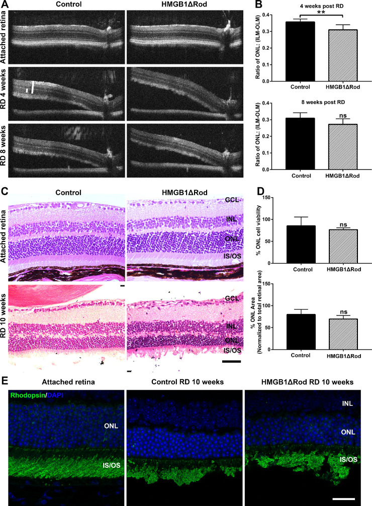
Figure 4.
Conditional knockout of HMGB1 from rods accelerates their degeneration during RD. (A) OCT images in attached and detached retinas at 4 and 8 weeks post-RD in HMGB1ΔRod and control mice. Due to the angle created by retinal detachment, a novel method was developed to measure the thickness of the retina to ensure that corresponding points on the retinas were measured (see Materials and Methods). (B) The thickness of ONL and the thickness from the ILM to the OLM were measured at 500 pixels away from the optic nerve head using ImageJ. The ONL/(ILM-OLM) ratio was calculated and graphed. (C) Hematoxylin and eosin staining of retinal sections was performed at 10 weeks post-RD in HMGB1ΔRod and control mice. (D) The nuclei count in the ONL in the detached portion of retinal sections was performed using ImageJ. The ONL area and the area between the ILM and OLM in the detached portion of the retina were also measured. The ONL/(ILM-OLM) area ratio was calculated. Normalized data were graphed with controls set as 100%. (E) Retinal sections of HMGB1ΔRod and control mice at 10 weeks post-RD were probed with antirhodopsin Ab. Nuclei were counterstained with DAPI. Scale bars: 50 µm. **P < 0.01 (n = 5, unpaired Student's t-test). ns, not significant.

