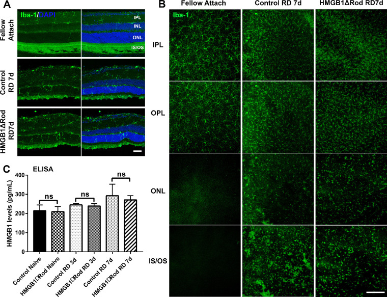
Figure 5.
Conditional knockout of HMGB1 in rods does not affect microglial activation or mobilization. (A) Retinas of HMGB1ΔRod and control mice were sectioned at a 30-µm thickness and probed with anti–Iba-1 antibody to view activation and mobilization of microglia/macrophage at 7 dprd. Nuclei were counterstained with DAPI. Scale bar: 50 µm. (B) Entire retinas of HMGB1ΔRod and control mice were probed with anti–Iba-1 antibody and flat-mounted for confocal imaging at 7 dprd. Multiple confocal images were Z-stacked to create merged images of different layers of the retina based on the DAPI staining. Scale bar: 200 µm. (C) Supernatants from eyecups were collected and the levels of HMGB1 in HMGB1ΔRod and control mice at 3 and 7 dprd were measured by ELISA. One-way ANOVA (n = 4). IPL, inner plexiform layer; IS/OS, inner and outer segment; ns, not significant; OPL, outer plexiform layer.

