Figure 6.
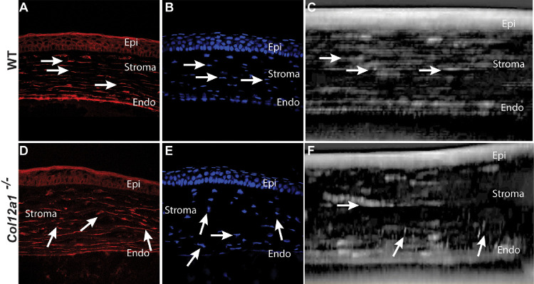
Disorganized keratocyte distribution and networks in tissue sections and reconstructed stromal images from in vivo confocal microscopy. (A) Actin filaments showed well-aligned keratocytes oriented in the interlamellar space. (D) In contrast, keratocyte organization and alignment were disrupted in the absence of collagen XII. (B) DAPI showed keratocyte nuclei horizontally aligned between stromal lamellae. (E) DAPI nuclear staining showed disorganized nuclei orientation and arrangement in Col12a1–/– corneas. (C) Reconstructed in vivo confocal two-dimensional image also shows well-aligned keratocytes in the horizontal plane. (F) In contrast, disrupted organization and alignment were noted in a representative in vivo confocal two-dimensional image in Col12a1–/– corneas.

