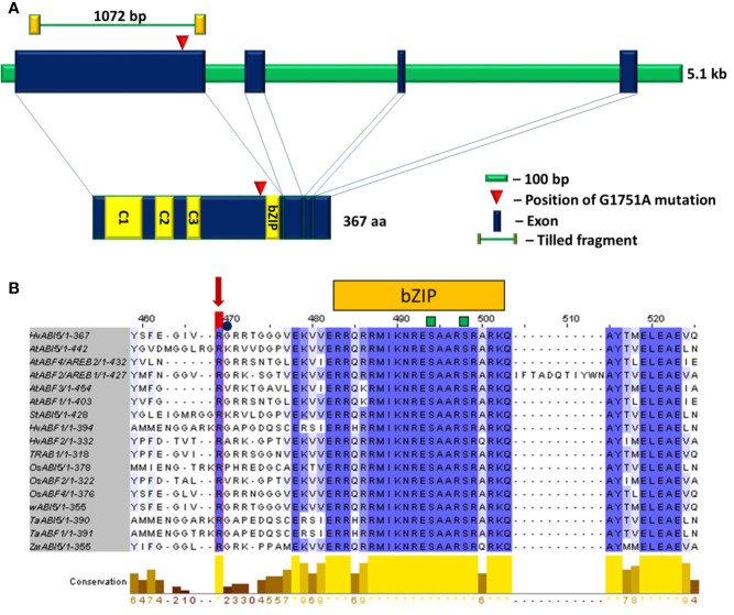
Figure 3.
(A) The structure of HvABI5 gene and HvABI5 protein with the position of hvabi5.d mutation indicated. C1, C2, C3—conserved charged domains, bZIP—basic leucine zipper domain. (B) An alignment of ABI5 and ABF proteins fragment comprising hvabi5.d mutation position. The changed arginine (R274) in hvabi5.d is marked by a red arrow. The square and circle mark phosphorylation and ubiquitination sites, respectively. Blue color indicates conservation of aligned position, yellow bars mark level of conservation. The numbering visible in alignment visualization refers to multi sequence alignment of 17 ABI5 and ABF protein sequences in dicot and monocot species. It does not indicate the position of amino acids substituted in the hvabi5.d mutant given in Tab.1.

