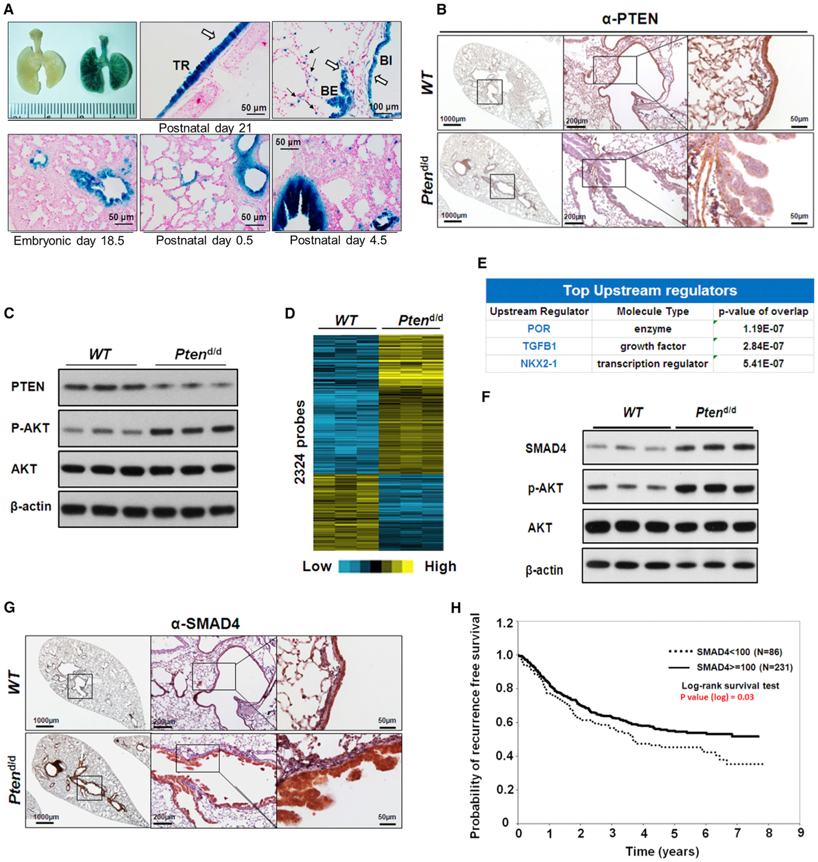
Figure 1. Deletion of Pten in Mouse Bronchial Epithelial Cells Results in Hyperplasia and Alters TGF-β Signaling.

(A) Representative whole-mount X-gal staining of mouse lungs in different stages. The blue X-gal staining showed CCSPiCre expressed in tracheal and bronchial airway epithelial cells in R26R (left) and CCSPiCre/R26R (right) mice (upper panel). The white arrows indicate the blue staining of X-gal in tracheal and bronchial airway epithelial cells and the black arrows indicate the staining of X-gal in the alveolar type II cells; trachea (TR), bronchi (BI), and bronchiole (BE).
(B) IHC staining of PTEN in lungs of 7-month-old WT and Ptend/d mice. PTEN staining is strongly present in the epithelium (brown staining) of WT mice and not in the epithelial cells of the Ptend/d mouse lung.
(C) WB analysis of PTEN and AKT expression and Akt phosphorylation (p-AKT) in lungs of 7-month-old mice. Increased p-AKT was observed in Ptend/d mouse lungs.
(D) The heatmap for the 1,847 differentially regulated genes (2,324 probes) in the microarray analysis of lungs from 7-month-old Ptend/d mice (n = 6) when compared to 7-month-old WT mice (n = 6).
(E) IPA of gene microarray data revealed top changed upstream regulators in Ptend/d mice, as compared to WT mice.
(F) WB analysis of SMAD4 protein expression in 7-month-old mouse lungs.
(G) IHC staining of SMAD4 (brown staining) in 7-month-old WT and Ptend/d mouse lungs.
(H) Kaplan-Meier plot of probability of recurrence-free survival based on the cytoplasmic SMAD4 protein expression index in lung cancer patients from SMAD4 tissue array data set.
