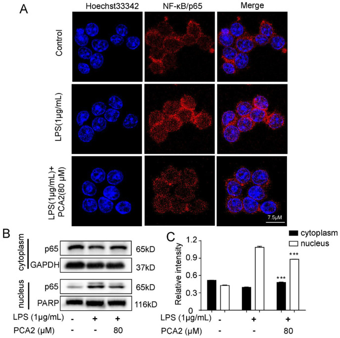
Fig 5. PCA2 blocked LPS-induced NF-κB/p65 translocation.
(A) The LPS-induced group cells were exposed to LPS for 2 h, while the experiment group cells were exposed to PCA2 (80 μM) for 1 h and cultured with LPS for another 1 h. Subsequently, cells incubated with NF-κB p65 antibody and secondary antibody for 1 h independently. Later, we used Hoechst 33342 to stain nuclei for a quarter. Images were taken with the confocal laser scanning microscopy. (B) The cell was exposed to PCA2 (80 μM) for 1 h and cultured with LPS for 2 h, except for the control group. The protein expressions of p65 were detected by western blotting (n = 3). (C) Statistical analysis of the p65 protein expression in the cytoplasm and the nucleus (n = 3). *** p < 0.001 vs. LPS group.

