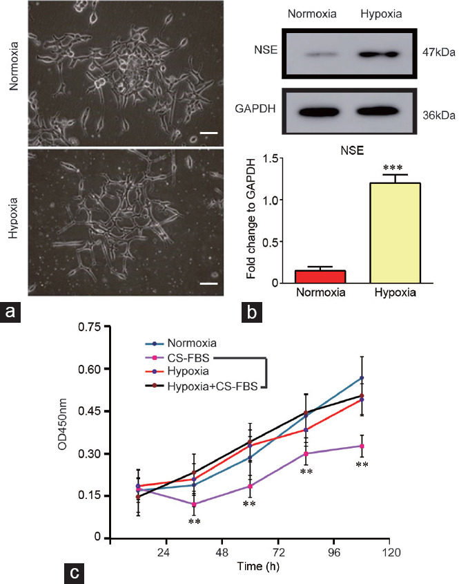
Figure 2.

(a) Representative images of LNCaP cells cultured under hypoxic and normoxic conditions for 14 days (×100, scale bars=10 μm). (b) Western blot assay showing the expression of NSE in LNCaP cells cultured under hypoxic conditions. The quantification analysis is presented in the lower panel. (c) Cell proliferation results of LNCaP cells and 14 days hypoxia-cultured LNCaP cells cultured in medium with or without DHT, as determined by a CCK8 assay. The experiments were performed in triplicate **P < 0.01 and ***P < 0.001. DHT: dihydrotestosterone; CS-FBS: charcoal-stripped fetal bovine serum; NSE: neuron-specific enolase; GAPDH: glyceraldehyde-phosphate dehydrogenase; OD: optical density; CCK8: cell counting kit 8.
