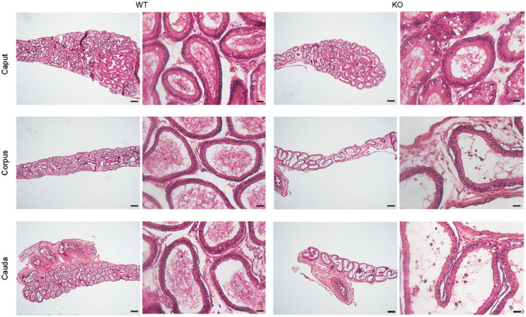
Figure 4.

Hematoxylin-and-eosin staining of the epididymal sections from 5-week-old Plag1 WT and KO mice. Scale bars represent 200 μm in low-magnification images (left column) and 20 μm in high-magnification images (right column). Plag1: pleomorphic adenoma gene 1; WT: wild-type; KO: knockout.
