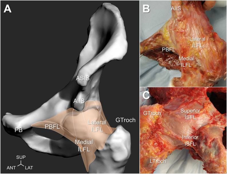
Fig. 1.
Anatomy of the capsular ligaments illustrated with a left-sided hip model in neutral position (Fig. 1-A) and showing the anterior view of a cadaveric hip specimen in external rotation (Fig. 1-B) and the posterior view of a cadaveric hip specimen in internal rotation (Fig. 1-C). The lateral and medial branches of the iliofemoral ligament (ILFL), pubofemoral ligament (PBFL), superior and inferior fibers of the ischiofemoral ligament (ISFL), anterior superior iliac spine (ASIS), anterior inferior iliac spine (AIIS), pubis (PB), and greater and lesser trochanters (GTroch and LTroch) are indicated. SUP = superior, ANT = anterior, and LAT = lateral.

