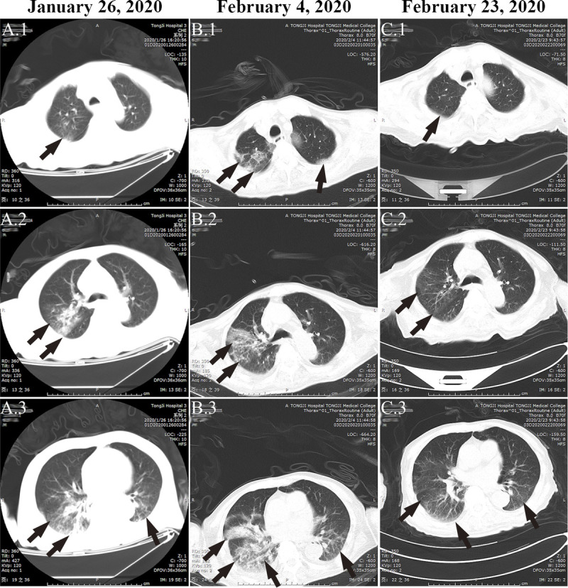
FIGURE 1.

The evolution process of COVID-19 pneumonia in case 1. For case 1, on the day of onset on January 26, 2020, CT images indicated pneumonia (as indicated by arrows), but there were no typical manifestations of peripheral subpleural ground-glass opacities (A.1–A.3). A rapid progression of pneumonia was observed on the CT image 9 days after onset (B.1–B.3). Computed tomography re-examination indicated significant improvement on February 23, 2020 (C.1–C.3).
