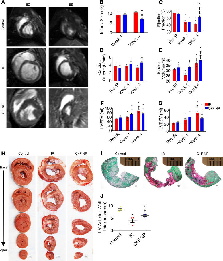
Figure 5. Assessment of LV morphology and function in a pig model of IR injury.
Cardiac MRI recordings in the experimental groups at end-diastole (ED) and end-systole (ES). At day 28, the CHIR + FGF1-NP-treated group revealed significant reduction in infarct size compared with the untreated IR group (B). On the contrary, EF (C), CO (D), and SV (E) were significantly greater in CHIR + FGF1-NP–treated groups than in untreated IR groups while LV end-diastolic volume and LV end-systolic volume were significantly lower (F and G). Data are given as means ± SEM. There were 4 animals per group. Statistical analysis: 2-way ANOVA with Dunn’s multiple comparisons test. *P < 0.01 vs. pre-IR; †P < 0.05 vs. week 1; ‡P < 0.05 vs. IR. Macroscopic areas of infarction/fibrosis/scar (H) at after-IR day 28 in serial transverse sections of fresh (scale bar: 1 cm) representative micrographs of Sirius Red/Fast Green histochemical staining, revealing areas of infarcted (red, nonviable) and noninfarcted (green, viable) zones (I) (scale bar: 1 mm) and quantification of left anterior wall thickness (J). Data are given as means ± SEM. There were 4 animals per group. Statistical analysis: 1-way ANOVA with Dunn’s multiple comparisons test. *P < 0.01 vs. control; †P < 0.05 vs. IR.

