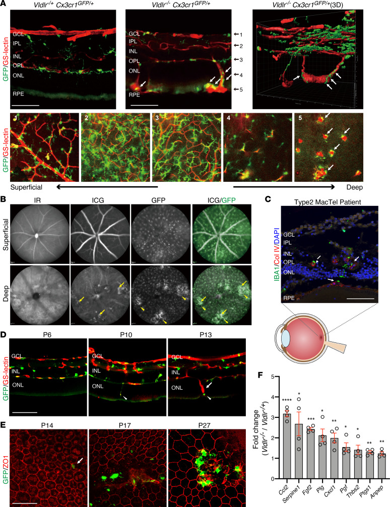
Figure 1. Retinal microglia or macrophages are associated with subretinal neovascular angiomas in Vldlr–/– mice.
(A) GS-lectin staining on cryosectioned Vldlr-/+; Cx3cr1GFP/+ and Vldlr–/–; Cx3cr1GFP/+ retinas at P27 (top) and flat-mounted Vldlr–/–; Cx3cr1GFP/+ retina in each retinal layer (bottom) indicate Cx3Cr1-GFP–positive microglia/macrophages (white arrows) in close proximity of abnormal angiomas in the outer nuclear layer and subretinal space in Vldlr–/–; Cx3cr1GFP/+ mice. (B) VEGF fundus autofluorescence by infrared reflectance (IR), indocyanine green angiography (ICG), and GFP in P40 Vldlr–/–; Cx3cr1GFP/+ mice showed that the pathological neovascular angiomas (yellow arrows) and the GFP-positive cells were aggregated close to the neovessels (yellow arrowheads). (C) IBA1-positive microglia/macrophages were localized in the outer nuclear layer and subretinal space in proximity of neovessels positive for collagen IV (Col IV; counterstained with DAPI, white arrows) in a retinal section of a macula from a patient with MacTel. (D) GS-lectin staining on cryosectioned P6, P10, or P13 Vldlr–/–; Cx3cr1GFP/+ retinas demonstrates that microglia (arrowheads) migrated into the subretinal space ahead of the neovessels (arrow). (E) Immunostaining for ZO1 in flat-mounted RPE at P14, P17, and P27 of Vldlr–/–; Cx3cr1GFP/+ mice. At P14, the first appearance of GFP-positive microglia/macrophages migrated into ZO1-positive RPE layer was observed (white arrow) with subsequent NV following. (F) Significantly upregulated genes between Vldlr–/– and Vldlr-/+ mice, as analyzed using a PCR array for angiogenesis, are shown (P < 0.05 and >1.5 fold change) The P values were calculated based on a Student’s t test of the replicate 2(- Delta Ct) values for each gene in the Vldlr-/ group and Vldlr-/+ groups. *P < 0.05, **P < 0.01, ***P < 0.001, ****P < 0.0001 (n = 4 each). GCL, ganglion cell layer; IPL, inner plexiform layer; INL, inner nuclear layer; OPL, outer plexiform layer; ONL, outer nuclear layer; RPE, retinal pigmented epithelium. Scale bars: 100 μm (A, C, and D); 50 μm (E).

