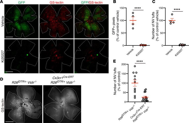
Figure 2. Microglia ablation prevents subretinal neovascularization in Vldlr–/– mice.
(A–C) Pharmacological microglia ablation using the CSF-1R inhibitor Ki 20227 in Vldlr–/–; Cx3cr1GFP/+ mice. (A) Subretinal neovascular (NV) tufts and GFP-positive microglia/macrophages were analyzed using GS-lectin staining in P17 Vldlr–/–; Cx3cr1GFP/+ mice treated with vehicle or Ki20227 from P10 to 16. Both (B) GFP-positive pixels and (C) the number of subretinal NV tufts were significantly reduced in Ki20227-treated mice retina. The P values were calculated using an unpaired 2-tailed t test (vehicle: n = 4, Ki20227: n = 6). (D and E) Genetic ablation of microglia in Vldlr–/– mice was achieved by crossing Cx3cr1Cre-ERT; Vldlr–/– mice and Rosa26iDTR/+mice. (D) The NV tufts in P23 Rosa26iDTR/+; Vldlr–/– and Cx3cr1 Cre-ERT; Rosa26iDTR/+; Vldlr–/– mice were analyzed by GS-lectin staining after 4-hydroxytamoxifen (4HT) treatment at P10 and P11, followed by P12-P14 diphtheria toxin (DT) treatment, demonstrating a marked reduction of NV tufts in Cx3cr1 Cre-ERT; Rosa26iDTR/+; Vldlr–/– mice (Rosa26iDTR/+; Vldlr–/–: n = 11, Cx3cr1 Cre-ERT; Rosa26iDTR/+; Vldlr–/–: n = 15) (E). Error bars indicate the mean ± SEM. The P values were calculated using an unpaired 2-tailed t test. ****P < 0.0001. Scale bars: 1 mm.

