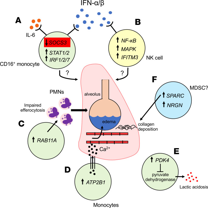
Figure 6. Potential immune mechanisms contributing to the development of ARDS.
(A) Monocyte subsets (particularly CD16+ monocytes) downregulate SOCS3 expression, which promotes type I IFN signaling through STAT and IRF pathways. Reduced SOCS3 levels may also modulate IL-6 signaling. (B) NK cells have increased expression of canonical activation signaling cascades including NF-κB, MAPK, and IFN-stimulated genes (IFITM3). (C) Monocytes with increased expression of RAB11A may impair neutrophil efferocytosis, leading to persistent alveolar inflammation. (D) Increased ATP2B1 is involved in calcium efflux, which could contribute to alveolar capillary leak and the development of noncardiogenic pulmonary edema. (E) Increased PDK4 could contribute to lactic acidosis through inhibition of pyruvate dehydrogenase. (F) Monocytes with increased SPARC and NRGN expression could be markers of myeloid-derived suppressor cells (MDSCs). SPARC is involved in collagen deposition, which is observed in the fibroproliferative stage of ARDS.

